MCR Diabetes & Eye Care · Kannur, Kerala
+91 9497 222 722
Abdominal Ultrasound: What the Scan Detects, How to Prepare, and What Findings Mean
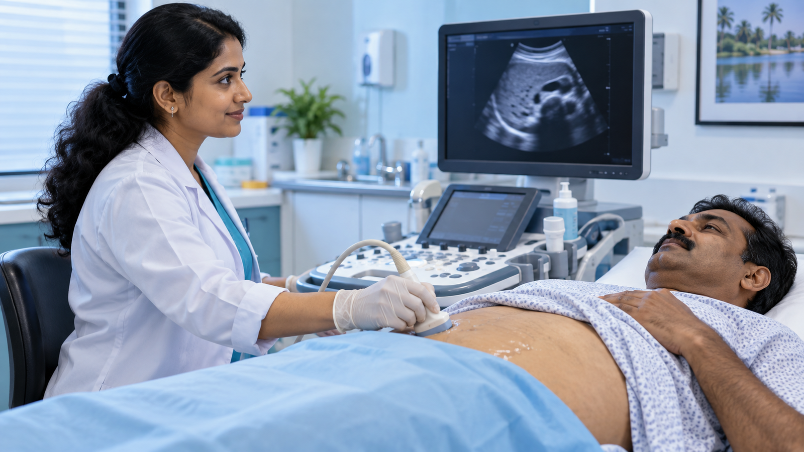
The abdominal ultrasound is among the most frequently ordered scans in India, and among the least explained. Patients arrive fasting, spend fifteen minutes with a probe on the abdomen, and leave with a report full of terms like “Grade 1 fatty liver,” “cortical echogenicity,” and “no free fluid.” Yet this scan is genuinely valuable — it can identify fatty liver, gallstones, some kidney stones, and structural abnormalities without ionising radiation, at modest cost.[1][2] Our specialist team at MCR Diabetes & Eye Care, Kannur, explains what it shows and what your report means.
6-8 hours
fasting required before the scan
No radiation
safe to repeat as often as needed
95%+
accuracy for detecting gallstones
₹800-2,000
typical cost in Kerala
Ultrasound uses high-frequency sound waves rather than radiation, making it safe to repeat as often as needed and safe in pregnancy. For metabolic patients it earns its place primarily as a fatty liver detector — and given that fatty liver affects up to 70% of type 2 diabetics, that is not a minor finding. See our fatty liver disease guide.
What the Scan Examines
| Organ | What It Can Detect | Reliability |
|---|---|---|
| Liver | Fatty liver, size, cysts, masses, cirrhosis changes | Good (misses mild fat) |
| Gallbladder | Stones, wall thickening, polyps | Excellent (95%+) |
| Bile ducts | Dilatation, obstruction | Good |
| Kidneys | Stones, cysts, hydronephrosis, cortical changes | Good |
| Bladder | Wall thickening, stones, residual urine | Good (needs full bladder) |
| Spleen | Size, focal lesions | Good |
| Pancreas | Masses, cysts, inflammation | Often obscured by gas |
| Aorta | Aneurysm | Good |
| Uterus / ovaries / prostate | Fibroids, cysts, enlargement | Good |
| Bowel | Very limited | Poor — not the right test |
Preparing for the Scan
- Fast for 6-8 hours before the scan. Food causes the gallbladder to contract, making stones and wall thickening difficult to see, and eating increases bowel gas which obscures deeper structures.
- Water is usually permitted — and if the pelvis or bladder is being examined, a full bladder is required. Ask when booking.
- Diabetics on medication: book an early morning slot and discuss with your doctor whether to delay your morning dose until after the scan. Never skip insulin without advice — see our insulin therapy guide.
- Avoid fizzy drinks the day before, as they increase gas
- Wear loose clothing with easy abdominal access
- Bring previous scans and reports — comparison is often more informative than the new scan alone
The scan itself takes 15-30 minutes, is painless apart from mild probe pressure, and involves no recovery time. Cost in Kerala typically runs ₹800-2,000.
Key fact: The pancreas is poorly visualised in a large proportion of abdominal ultrasounds because overlying bowel gas obscures it. A normal-looking pancreas on ultrasound does not exclude pancreatic disease — a common and important misunderstanding.
Common Findings Explained
Fatty Liver (Hepatic Steatosis)
Reported in grades 1 to 3 based on how bright the liver appears compared with the kidney. Grade 1 is mild and typically fully reversible; Grade 3 indicates substantial fat accumulation. Important limitation: ultrasound reliably detects moderate to severe fatty liver but frequently misses mild disease, and it cannot distinguish simple fat from inflammation or fibrosis. A FibroScan or blood-based fibrosis score adds that information.
Gallstones (Cholelithiasis)
Ultrasound is the gold standard here, with over 95% accuracy. Many gallstones are silent and found incidentally — these usually need no treatment. Stones causing pain, or associated with gallbladder wall thickening or bile duct dilatation, warrant surgical referral. Diabetics have a somewhat higher risk of complications from gallstone disease.
Renal Calculi (Kidney Stones)
Ultrasound detects most kidney stones and shows hydronephrosis (backpressure swelling) well. It is less reliable for small stones in the ureter, where CT is preferred.
Renal Cortical Echogenicity
Increased brightness of the kidney cortex suggests chronic kidney disease. In a diabetic, this finding should always prompt urine ACR and eGFR testing — see our diabetic nephropathy guide.
Simple Cysts
Very common in liver and kidneys, almost always harmless, and usually need no follow-up. Complex cysts with septations or solid components require further imaging.
Enlarged Prostate
Often reported with post-void residual urine volume. Common with age; treatment depends on symptoms rather than size alone.
Hepatomegaly or Splenomegaly
Enlargement of liver or spleen. Causes range from fatty liver and alcohol to infections and blood disorders, so the finding directs further testing rather than providing a diagnosis.
⚠ Important: A normal abdominal ultrasound is not a clean bill of abdominal health. It cannot assess the bowel, frequently misses mild fatty liver, and cannot grade liver fibrosis. Persistent symptoms deserve further investigation regardless of a normal scan result.
What Ultrasound Cannot Do Well
- The pancreas is often poorly visualised — overlying bowel gas obscures it in a large proportion of scans. A normal-appearing pancreas on ultrasound does not exclude pancreatic disease.
- Bowel pathology is largely invisible — ultrasound cannot reliably assess the intestines or diagnose colorectal disease
- Mild fatty liver may be missed
- It cannot reliably grade liver fibrosis — additional assessment, which may include elastography and blood-based fibrosis scores, is often needed.[4]
- Obesity and bowel gas reduce image quality substantially
- It is operator-dependent — the skill of the person scanning materially affects what is found
These limitations matter because a “normal ultrasound” is often taken as a clean bill of abdominal health, which it is not. Persistent symptoms deserve further investigation regardless of a normal scan.
Metabolic Screening Including Ultrasound
Ultrasound interpreted alongside liver enzymes, glucose, lipids, and kidney function — not as an isolated report.
Who Should Have One
- Anyone with type 2 diabetes, obesity, or metabolic syndrome — primarily for fatty liver screening. See our metabolic syndrome guide.
- Raised liver enzymes on blood tests
- Persistent abdominal pain, particularly upper right or upper left
- Unexplained weight loss
- Suspected gallstones — recurrent pain after fatty meals
- Blood in urine, flank pain, or recurrent urinary infections
- Abnormal kidney function tests
- As part of a comprehensive health check — see our annual check-up guide
Reading Your Report Sensibly
- Incidental findings are extremely common and mostly benign — simple cysts, small haemangiomas, mild prostate enlargement
- Grades and measurements are approximate, and can vary between operators and machines
- Comparison with previous scans is often the most useful information — keep your old reports
- The report describes; it does not diagnose. Interpretation requires your clinical picture and blood results alongside it.
Abdominal Ultrasound at a Glance
| Aspect | Key Point |
|---|---|
| Preparation | Fast 6-8 hours; full bladder if pelvis included |
| Duration | 15-30 minutes, painless |
| Radiation | None — safe in pregnancy and repeatable |
| Best for | Gallstones, fatty liver, kidney stones, organ size |
| Poor for | Pancreas, bowel, mild fatty liver, fibrosis grading |
| Fatty liver grading | Grade 1-3 by brightness vs kidney |
| Adds fibrosis info | FibroScan or FIB-4 blood score |
| Diabetics on medication | Book early slot; ask about morning dose timing |
| Bring | Previous scans — comparison is often most useful |
Frequently Asked Questions
Below are the questions our patients ask most often. If you have additional questions, our specialist team at MCR Diabetes & Eye Care, Kannur, is always available to help.
Why do I have to fast for the scan?
Food makes the gallbladder contract, hiding stones and wall changes, and eating increases bowel gas which obscures deeper structures like the pancreas. Six to eight hours of fasting gives much clearer images.
I have diabetes — is fasting safe?
Book an early morning slot to minimise the fasting window, and discuss with your doctor whether to delay your morning medication until after the scan. Never skip insulin without advice, and carry fast-acting glucose with you.
What does ‘Grade 1 fatty liver’ actually mean?
It indicates mild fat accumulation in the liver, based on how bright the liver appears compared with the kidney. Grade 1 is typically fully reversible with weight loss and dietary change — treat it as an early warning rather than a diagnosis to ignore.
My report mentions a simple cyst. Should I be worried?
Simple cysts in the liver or kidneys are very common and almost always harmless, usually requiring no follow-up. Complex cysts with septations or solid components are different and warrant further imaging.
Can ultrasound detect cancer?
It can detect many masses and abnormalities that prompt further investigation, but it cannot definitively diagnose cancer and misses some tumours entirely — particularly in the pancreas and bowel. CT, MRI, or endoscopy are used when cancer is suspected.
Is ultrasound safe to repeat frequently?
Yes. It uses sound waves rather than ionising radiation, so there is no cumulative dose concern. It can be repeated as often as clinically useful, including during pregnancy.
Why did two scans give different fatty liver grades?
Ultrasound is operator-dependent and grading is subjective, so mild differences between operators and machines are common. This is why comparison with previous reports, and correlation with blood tests, matters more than a single grade.
Final Takeaway: A Useful Scan, Read in Context
For diabetics and anyone with metabolic risk, an abdominal ultrasound is inexpensive, safe, and frequently the first test to reveal fatty liver — a finding with real consequences for cardiovascular and liver health. Its value depends on preparing properly, understanding its blind spots, and having the report interpreted alongside your blood work rather than in isolation.
At MCR Diabetes & Eye Care, Kannur, abdominal ultrasound forms part of our comprehensive metabolic screening, interpreted together with liver enzymes, glucose, lipids, and kidney function. If you have diabetes or metabolic syndrome and have never had one, book a screening today.
You May Also Find Helpful
- Fatty Liver Disease (NAFLD) in Indians
- Metabolic Syndrome: The Five Warning Signs
- Kidney Function Tests in Diabetes
- Annual Health Check-Up Guide for Indians
Evidence note: Ultrasound is recommended when gallstone disease is suspected, but it has important limits. In adults with suspected acute renal colic, NICE recommends low-dose non-contrast CT as the usual first-line test unless pregnancy makes ultrasound preferable. Conventional ultrasound can detect liver fat but does not by itself stage the severity of liver disease.[2][3][4]
Sources and Further Reading
- American Institute of Ultrasound in Medicine. Practice parameter for abdominal and retroperitoneal ultrasound.
- NICE. Gallstone disease: diagnosis and management (CG188).
- NICE. Renal and ureteric stones: assessment and management (NG118).
- AASLD practice guideline on imaging-based assessment of hepatic steatosis and fibrosis.